
Fish in a recirculating system live close together in treated water. A parasite, a bacterial infection, or a stretch of poor water quality can move through a tank faster than it would in a thinly stocked pond. Early detection is therefore not a luxury. It is part of ordinary husbandry. Microscopy remains one of the most direct ways to see what is on the gill, the skin, or in the water, and new instruments try to make that look faster and easier to record.
This article explains why health work belongs beside water-quality work in a recirculating workshop, what a microscope can and cannot show, and how Qiuhao 5 and Qiuhao 8 were publicly presented in September 2026. Company-stated identification figures are labelled as such. No unverified accuracy claim is offered as a certified result, and the WeChat mini-program associated with the instruments is described as a China offering only.
Why recirculating systems make health visible sooner
Density is the first reason. A recirculating tank is usually stocked more heavily than an extensive pond, because the cost of the building and the treatment loop has to be carried by more fish. At higher density, a pathogen that finds a host has more neighbours to reach. Stress from crowding, from a nitrite spike, or from a sudden temperature change also makes fish less able to keep parasites and bacteria in check. The same water reuse that saves water can, if hygiene is poor, recirculate pathogens as efficiently as it recirculates oxygen.
The second reason is observation. In a clear tank, under a roof, a skilled technician can see flashing, clamped fins, lethargy, unusual swimming, or a change in feeding within a single round. In a turbid pond those signs are easier to miss until mortality has already begun. A demonstration workshop should therefore be a place where staff are in the habit of looking, not only of reading a screen.
The third reason is the cost of a late response. In RAS, a treatment that requires a large water exchange is expensive, and some medications interfere with the biological filter. Preventing a problem, or catching it when only a few fish are affected, is better than trying to medicate a whole loop after the filter has been stressed. That is an argument for early detection, and it is also an argument for biosecurity: clean incoming stock, controlled access, separate nets, and a place to isolate sick fish.
Health work does not replace water-quality work. Many “disease” events begin as a water-quality event. Ammonia, nitrite, low oxygen, or a rapid pH change can damage gills and open the way for parasites and bacteria. A microscope slide that shows a parasite should always be read beside the week’s water log. The two records explain each other.
What microscopy can show, and what it cannot
A light microscope, used on a wet mount of skin mucus, a gill clip, or a water drop, can reveal many of the protozoan and metazoan parasites that trouble farmed fish, and it can show some algal cells that affect ponds and sometimes indoor systems. A competent operator looks at movement, shape, attachment, and number, and compares what is seen with the condition of the fish. The method is old, inexpensive in principle, and still central to farm and laboratory practice around the world.
What microscopy does not do, by itself, is name every bacterium, virus, or nutritional problem. Many bacterial diagnoses need culture or molecular methods. Viruses are not visible on a standard farm wet mount in the way a trichodinid or a gyrodactylid may be. Poor growth may have more to do with feed, temperature, or chronic stress than with a parasite that happens to be present in small numbers. A microscope is a powerful first instrument. It is not a complete hospital.
Skill still matters. Preparing a thin, fresh slide, using the right magnification, and knowing what a normal gill looks like are learned habits. A device that photographs a slide and suggests a name can help a less experienced user, or speed a busy laboratory, but it does not remove the need for judgement. False confidence is a risk with any identification aid, automated or not. The responsible use of such a tool is to support a decision, and to keep a record, not to replace a person who can still look at the fish.
That is the context in which a company might offer an “AI microscope”: as a way to standardise the first look, to store images, and to draft a report, especially where experienced microscopists are scarce. Whether a given instrument does that well is a question for demonstration, for comparison with a trained eye, and, where claims are numerical, for evidence that can be inspected. This site does not treat a press description as a substitute for that inspection.
Qiuhao 5 and Qiuhao 8, as they were publicly presented
On 10 September 2026, at the 12th Guangzhou International Fishery Expo, Guangzhou Nansha Zhihui Agricultural Technology Co., Ltd. launched two instruments, Qiuhao 5 and Qiuhao 8, for aquatic disease microscopy and identification support. Deputy General Manager Lei Lei spoke at the launch. Press reports described the products as tools with which a user examines a sample and the system supports identification and a suggested course of action. Domestic dealer recruitment was announced the same day.
The announced positioning of the two models is straightforward. Qiuhao 8 was described for research institutes, large farms, and professional testing laboratories — settings that need a higher standard of microscopic imaging. Qiuhao 5 was described for smaller farms, grassroots aquatic pharmacies, and front-line practitioners. Those are marketing positions published at a launch. They are not, in the public record used for this site, a full optical specification sheet, a price list, or an international certification.
A disease-recognition WeChat mini-program has been offered free of charge in China, including for users who do not purchase a microscope. That offering is a China-channel service. It is not presented here as a product promise for overseas buyers. A Gulf or Southeast Asian reader who is interested in microscopy support should treat the mini-program as a domestic Chinese channel, and should ask, on a visit, what image and report workflow could actually be used in their own regulatory and language setting.
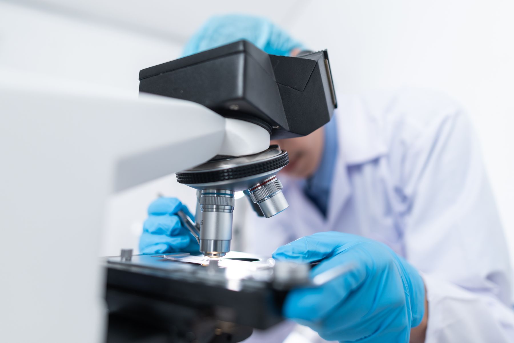
No CE mark or other international product certification is listed on this site for Qiuhao, because none has been publicly verified for these instruments. Official photographs of the devices will be added when the company can supply images it has the right to use. Until then, this article uses an illustrative laboratory photograph and says plainly that it is not a picture of Qiuhao.
Company-stated identification figures, labelled as such
A group party-committee article described the underlying identification work in these terms: more than 70,000 disease-sample images, more than 140,000 annotation boxes, and 71 common parasites and algae identifiable, with stated accuracy bands of more than 90 percent for 20 types, more than 80 percent for 24 types, and more than 70 percent for 18 types. The same article said devices were already in use at some aquatic hospitals and fish-disease testing centers, and that the equipment had completed design finalization and was entering launch preparation.
Those numbers come from a company-affiliated article. They have not been independently certified in the public record used to prepare this site. They should be read as company-stated figures. A serious buyer is entitled to ask how the test set was built, how “accuracy” was defined, whether the 71 types include the parasites that matter on their own farm, and how the system behaves on a poor slide or an unfamiliar species. Those questions are not hostile. They are how identification tools are properly bought.
Press at the launch also reported cooperation with named Chinese testing and technology firms, and a collaboration on staining methods for AI microscopic diagnosis with Xu Liwen of the South China Sea Fisheries Research Institute, Chinese Academy of Fishery Sciences. Those names are recorded on this site as reported cooperation, not as overseas deliveries or as third-party certificates of accuracy.
Daily health practice around the microscope
A workshop that uses a microscope well has a routine. A small number of fish from a tank that is not feeding well, or that shows flashing or mortality, are examined the same day. Gill and skin samples are taken humanely and viewed fresh. Findings are written down, with the tank number, the water-quality readings, and any treatment. Follow-up slides are taken after an intervention, so that staff can see whether the picture has changed. Dead fish are not left in the tank. Nets and buckets are not shared casually between a sick tank and a healthy one.
Incoming stock is a special case. A recirculating system is a poor place to discover that a delivery brought in a parasite. Quarantine tanks, a first microscopic screen, and a period of observation before fish join the main loop are ordinary biosecurity, not an advanced extra. A visitor can reasonably ask how incoming animals are handled in Nansha, and what happens when a slide is positive.
Feed and density belong in the same conversation. Overfeeding fouls water and stresses fish. Underfeeding also stresses fish. A health record that never mentions feed conversion or leftover pellets is an incomplete record. The management platform described by the company includes production logs as well as water-quality modules. Linking those logs to health events is one of the more useful things a digital system can do, provided someone still looks at the fish.
Treatments, when they are used, must be compatible with the biological filter and with any plants in an aquaponic loop, and they must be legal in the market where the fish will be sold. A suggested course of action from an identification tool is a starting point for a professional decision, not a prescription that can be followed blindly across borders. Veterinary and regulatory advice in the buyer’s own country remains necessary.
What a visitor should ask
If microscopy is part of the reason for a visit to Nansha, the useful questions are concrete. May I see how a slide is prepared? How is an image stored, and who can review it later? Which parasites does the staff most often encounter in this workshop, and how do they distinguish a true problem from a low background presence? How are company-stated identification figures explained, and what would a trial on the visitor’s own samples involve? What support, language, and spare parts could be discussed for an instrument used outside China?
Those questions can be asked without any claim that Qiuhao has already been certified or sold in the visitor’s market. Early detection is a general need in recirculating aquaculture. The instruments launched in 2026 are one company’s publicly announced attempt to support that need. A demonstration on the floor, with honest captions and conservative claims, is the right way to take the conversation further.
Health in RAS is a daily partnership among water chemistry, husbandry, biosecurity, and the ability to see small things before they become large ones. A microscope, used with judgement, remains central to that partnership. New tools may help more people to use it. They do not remove the need to look, to record, and to act.
A closer look at what is commonly sought on a slide
The parasites and irritants that a farm microscopist most often hopes to confirm or rule out are not mysterious, even if their names are Latin. Motile protozoans on the skin or gill, sessile ciliates, monogenean flatworms attached to gill filaments, and, in some waters, fungal hyphae or abundant algal cells, make up a large share of first-look findings. The operator is not completing a taxonomic monograph. The operator is asking whether what is present is enough, in this water and at this density, to explain the behaviour of the fish.
Numbers matter as much as names. A single organism on one slide is not the same as a heavy infestation on several fish. A written record that says only “parasite present” is weaker than one that notes which tissue, how many fields were viewed, and whether the finding was consistent across fish. If an identification tool proposes a name, the same discipline should still apply. A name without a sense of intensity, and without the week’s ammonia and oxygen readings, will mislead a later reader.
Sampling should be gentle and limited. A gill clip or a mucus scrape is a diagnostic act, not a casual souvenir. Fish that are already weak can be killed by clumsy handling. Staff should know how many animals they are allowed to sample from a given tank, how those animals are chosen, and how they are returned or humanely killed. A demonstration workshop can show this routine. A visitor should not be invited to handle fish without guidance.
Water samples have a place beside fish samples. A drop of tank water can show an algal bloom, a swarm of free-living ciliates, or a load of organic debris that helps to explain gill irritation. It cannot replace a look at the fish. The two views together are better than either alone. In an aquaponic workshop, a look at the plant side — roots, film, unusual odours — can also hint at a water-quality story that will later appear on a gill.
Biosecurity is part of early detection
Detection is late if the farm has already invited a problem in. Incoming stock, incoming water, staff movements, shared equipment, and visitors are the usual routes. A recirculating hall with a single poorly controlled door is not “high biosecurity” merely because it has a roof. Simple measures — a visitor log, dedicated footwear, hand cleaning, a rule about who may enter which room, and a quarantine line for new fish — do more than a poster on the wall.
When a problem is found, the next hour matters. Affected tanks should not share nets with unaffected ones. Mortality should be recorded by tank and by time, not remembered as a vague bad week. If a treatment is planned, the biological filter and any plants must be considered, and the legal position in the market of sale must be considered. Isolation is not always possible in a compact workshop, but a written plan for the attempt is better than improvisation in front of rising losses.
Training is the slow part of this work. A new technician can learn to cut a slide in a day and still need months to trust what they see. An instrument that stores images can help a senior colleague to review a junior’s work at a distance, which is one of the more honest uses of a digital microscope workflow. Remote review, however, still needs a person at both ends and a network that actually works. It is a support model to be discussed, not a claim of a worldwide duty diagnostician on every farm.